We use cookies to make your experience better. To comply with the new e-Privacy directive, we need to ask for your consent to set the cookies. Learn more.
ELISpot & Fluorospot Products
From research to discovery
Step 1
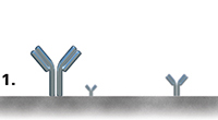
The cytokine-specific capture antibody is coated onto the PVDF-bottomed 96-well microtitre plate (not required for the pre-coated plates).
Step 2
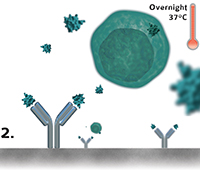
The cell suspension is added to the wells and incubated overnight at 37°C in a CO2 incubator, allowing the coated antibody to capture the target cytokine while being secreted.
Step 3
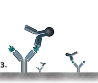
The biotinylated cytokine-specific detection antibody binds to the capture cytokine.
Step 4
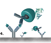
The alkaline phosphatase-conjugated streptavidin binds specifically to biotin.
Step 5
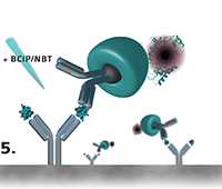
The BCIP/NBT substrate is added and turns into coloured spots wich can be quandtified either by an appropriate analysis software or manually by microscopy.
Caption
Capture /
Detection
antibody
Cytokine
Cytokine-
secreting
cell

Biotin

Streptavidin
Alkaline
phosphatase